 Sekar Ramachandran, Cornell University
Sekar Ramachandran, Cornell University
Thursday, September 6
4:00 p.m.
CNS 112
Refreshments served at 3:45, 1st floor CNS foyer. Bring your mug. Reduce, Reuse, Recycle.
Individuals with disabilities requiring accommodations should contact Nancy Pierce at npierce@ithaca.edu or (607) 274-3161. We ask that requests for accommodations be made as soon as possible.
Abstract:
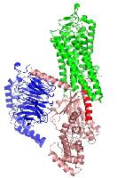
The phototransduction cascade in the mammalian rod cell provides an excellent model system to understand the mechanism of activation of G-proteins by their cognate G-protein coupled receptor (GPCR). Rhodopsin, with seven transmembrane helices, is activated by photons. Transducin, the G-protein that couples to rhodopsin, is made of three subunits designated aT, b1 and g1. The b1 and g1 are constitutively associated as a complex (bgT). The a-subunit of transducin (aT) binds guanine nucleotides with high affinity and in its guanosine diphosphate (GDP) bound form exists as a complex with bgT. Light triggers the activation of rhodopsin resulting in the release of the bound GDP from the aT subunit. The rate of GDP release from the aT subunit is extremely slow, on the order of a few hours, in the absence of light-activated rhodopsin. Activation of rhodopsin by light causes the release of GDP in a few seconds and the binding of guanosine triphosphate (GTP) to the aT subunit. The GTP-bound aT subunit is capable of activating its downstream target, cGMP phosphodiesterase (PDE). PDE catalyzes the hydrolysis of cGMP to GMP resulting in the closure of cGMP-gated ion channels. This triggers the release of a neurotransmitter and signaling to the brain via the optic nerve. I will discuss the use of in vitro biochemical and biophysical techniques to obtain a detailed understanding of the first step in vision, the catalysis of the release of GDP from transducin by rhodopsin.
https://www.ithaca.edu/intercom/article.php/20120828120731406